Blastomycosis
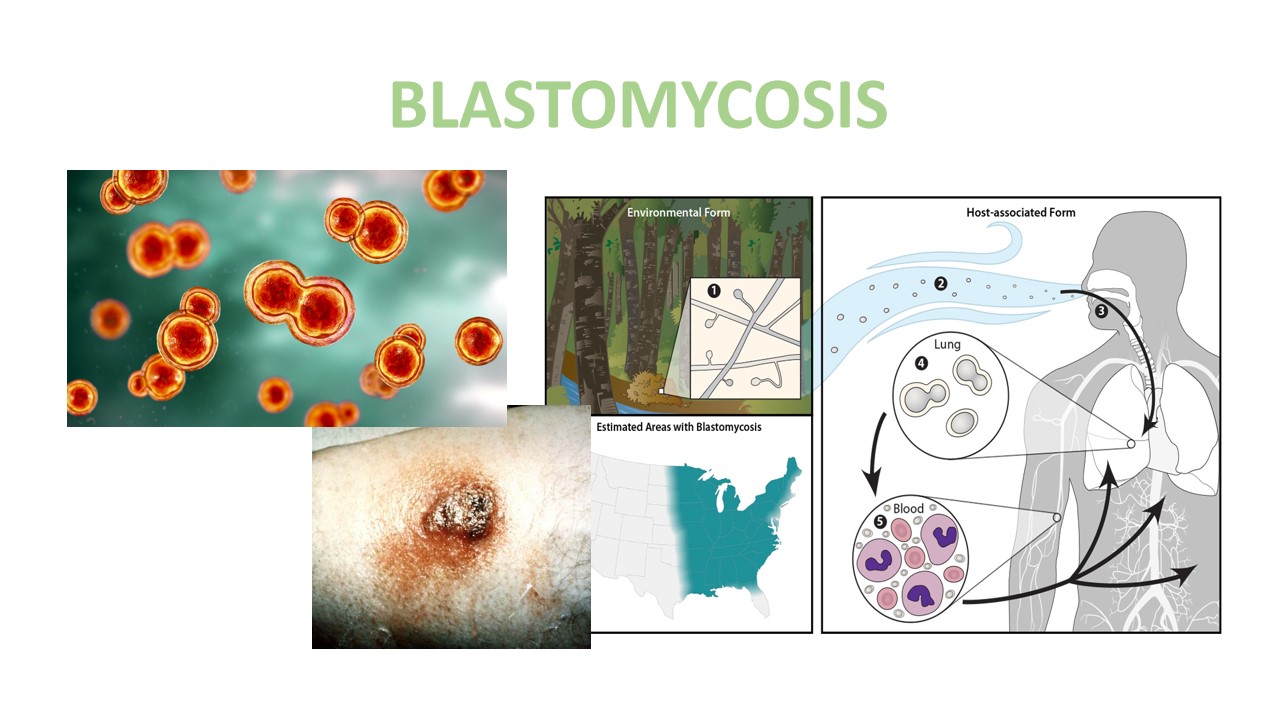
Blastomycosis is also called Gilchrist disease.Blastomycosis is a fungal disease. Blastomycosis is a pulmonary disease caused by inhaling spores of the dimorphic fungus Blastomyces dermatitidisor blastomyces gilchristi fungus. Blastomyces is naturally occurring in the environment, in the soil, organic composite matter such as wood and leaves. People may risk to blastomycosis when breathing the microscopic fungal spore from the air. Its mostly affect to immunocompromised person.
Blastomyces are naturally present in the environment in the form of mold. They produce tiny fungal spores that can’t see through naked eye. When environmental changes take place, it makes the soil and plants disturbed and release the small spores, then these tiny spores are mixed into the air. When people(weak immune system)breathe these spores from the air .They may risk to get the blastomycosis. In which the lungs are mostly affected. In the lungs, the spores develop into yeast and cause lung dysfunction . The spore can stay in the lung and can move through the blood stream to the other part of the body, such as skin, joints, organs and central nerve system. Blastomycosis is not contagious, it is not spread from person to person.
Symptoms
- Fever
- Cough
- Shortness of breath
- Night sweats
- Muscle aches or joint pain
- Weight loss
- Chest, rib, or back pain
- Fatigue (extreme tiredness)
Cause/Risk factors
- digging
- clearing wood
- excavation
- construction
Diagnosis
Cytology department
Culture cytology smears method is use to diagnosed the blastomycosis.The cytology specimens such as sputum are smeared on the slide with Periodic acid-Schiff(PAS) stain and observed under the microscope. The suspected orgnism are showed under the microscope.

sputum cytology
Microbiology department
The fungal culture is growing on the media plate. The fungal grown on the media plate takes 4-5 weeks. Take a small portion on a slide and observe under the microscope.

Microbiology examination
Histology department
Blastomycosis is diagnosed by a specimen like blood, urine and a biopsy of the infected area. The histological examination(tissue biopsy) is perform along with the microscope. In the histologycal examination, there are use specific stain such as hematoxylin, eosin, Gomori’smethenamine silver stain and periodic acid-Schiff(PAS) hematoxylin and eosin stain are use to detect the blastomycosis. Hematoxylin stain make the nuclei bluish-purple in colour and eosin stain make the rest of cellular component in pink to red . Gomori’s methenamine silver stain make the yeast cell wall deep black, it is use when background is green. Periodic acid-Schiff(PAS) stain makes the yeast cells red when they have a pink background or green light. Before the staining process, the pathologist performs the fixation, dehydration, embedding,mounting as per the laboratory process. After that, observed under a microscope.

Blastomycosis with Lung biopsy
Reference:
- https://www.cdc.gov/fungal/diseases/blastomycosis/index.html
-
https://universe84a.com/blastomyces-dermatitidis-general


0 comments