Cellulitis Disease
Cellulitis is one of the bacterial skin diseases. The skin acts as a protective layer against the normal skin flora and other microbial pathogens from reaching the subcutaneous tissue and lymphatic systems. When injuries happen in the skin, the normal skin flora and other pathogens invade the dermis and subcutaneous tissue. The entry of these bacteria, below the skin surface that are affecting the dermis subcutaneous tissue causing the cellulitis. The most frequent cause of cellulitis is an infection with group A beta-hemolytic streptococci(Streptococcus pyogenes).
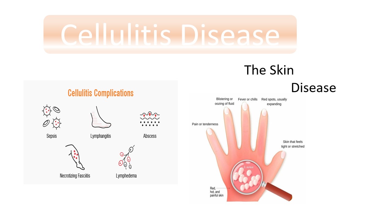
Cellulitis can affect any area of the body, however it most frequently affects the face and legs. Usually, the leg is mostly infected. The symptoms likely swelling and redness in the infected area. The skin may be puffy, and the boundaries of the redness are typically not sharp. The most often implicated microorganisms are Staphylococcus aureus and streptococci.Unlike cellulitis, erysipelas is a bacterial infection that affects the skin's outermost layers. It is often recognize as a red spot with distinct borders and is accompanied by a fever.
Symptoms
- Fever with chills and sweating
- Fatigue
- Pain or tenderness in the affected area
- Skin redness or inflammation that gets bigger as the infection spreads
- Skin sore or rash
- Tight, glossy, stretched appearance of the skin
- Warm skin in the area of redness
- Muscle aches and joint stiffness
- Nausea and vomiting
Causes/Risk factors
- Cracks or peeling skin between the toes
- History of peripheral vascular disease
- Injury or trauma with a break in the skin (skin wounds)
- Insect bites and stings, animal bites, or human bites
- Microoranism likeStreptococci and Staphylococcus aureus
Diagnosis
Microbiology Department: skin aspirate sample used to cultivate the bacteria on the media plate. After the colonies appeared, then these colonies were observed under the microscope by after performing the Gram staining technique.

Streptococcus pyogenes Gram positive bacteria observed under the microscope
Histology Department: The Histology section of the infected tissue can be used to diagnose cellulitis. The skin biopsy used as a specimen.The Gram stain is primarily used to distinguish between Gram-positive and Gram-negative bacteria based on their cell wall characteristics. In cases, any suspicion of bacterial infection causing cellulitis, a gram stain on the skin biopsy sample may reveal the presence of bacteria and their identification.

The pathogen observed under the microscope
Reference:
- https://www.mayoclinic.org/diseases-conditions/celiac-disease/symptoms-causes/syc-20352220
- https://www.hopkinsmedicine.org/health/conditions-and-diseases/celluliti
- https://en.wikipedia.org/wiki/Cellulitis


0 comments