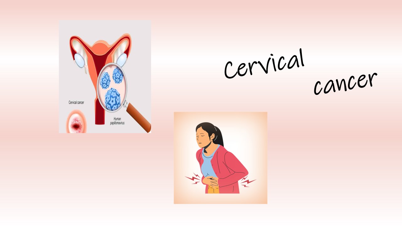
Cervical cancer
Cervical cancer is related to the female reproductive organ.It is cancer of the cervix. The Cervix is located at the lower end of the uterus at the junction of the vagina. When abnormal and uncontrolled growth of cells or changes happens in the cervix, it turns into cervix cancer. Cervix cancer can affect the deeper tissue of the cervix and spread to the other parts of the body, including the lungs, liver, bladder, vagina and rectum. Human papillomavirus (HPV) is responsible to cause cervix cancer. It occurs most often in people over the age of 30. People may have get viruses while having intercourse with an infected partner or cancer occurs when abnormal growth of cells or changes happen in the cervix due to radiation, changes in DNA and environmental changes.
There are three types of cervical cancer, such as squamous cell carcinoma, Adenocarcinoma and mixed carcinoma.The Squamous cells are thin, flat, skin-like cells, that lines are the surface of the cervix. This type of cancer develops where the cervix joint and cervix canal are connected. This is also called the transformation zone. Adenocarcinoma is less common, inside the cervix canal there are gland cells. These cells produce mucus sticky fluid to protect the cervix, womb, ovaries and Fallopian tubes from bacteria and infection. When the changes occur in gland cells, it can turn into adenocarcinoma. The screening of these types of cancers is difficult because it develops in the cervix canal. Mixed carcinoma include both squamous and adenocarcinoma. In which cancer cells from squamous cell and gland cells have to be infected. .Cervical cancer grows slowly, so that there is time to screen and treat them. The screening done by a pap test.
Symptoms
- pelvic pain or pain during sex
- Menstrual bleeding is heavier and lasts longer than usual.
- Watery, bloody vaginal discharge that may be heavy and have a foul odor.
- Vaginal bleeding after intercourse, between menstrual periods or after menopause.
- Pain in the abdomen
- Dull backache or swelling in your legs
cause/ risk factor
- Human papillomavirus (HPV) infection
- Oral contraceptives
- Exposure to diethylstilbestrol (DES
- Immune system deficiency
- Smoking tobacco
- obese, which may make it harder to diagnose cervical cancer
Diagnosis:
Cytology Examination: (Pep test)
Cervical cancer is diagnosed by a pap smear. This test is used to examine females' health conditions.After the sample was obtained,then make a smear and perform a microscope examination. Before that, the slide has to be stained with pap stain and reagent. In this procedure, the smear slide fixed in 95% alcohol.take a slide to hydrate with water. Rinse it in D/W.Stain it with hematoxylin for 2-3 Min's.After that, rinse with D/W. Dip it for 3 Min's in hydrochloric acid. Wash it with running water 2-3 Min . Then stain with cytologic stain, rinse with alcohol.Stain with EA-36 FOR 2 Min's, rinse with alcohol.clear xylene with alcohol. Mount it and observe under a microscope.

Reference:
- https://www.cancer.gov/types/cervical
-
https://www.mayoclinic.org/diseases-conditions/cervical-cancer/diagnosis-treatment/drc-20352506


0 comments