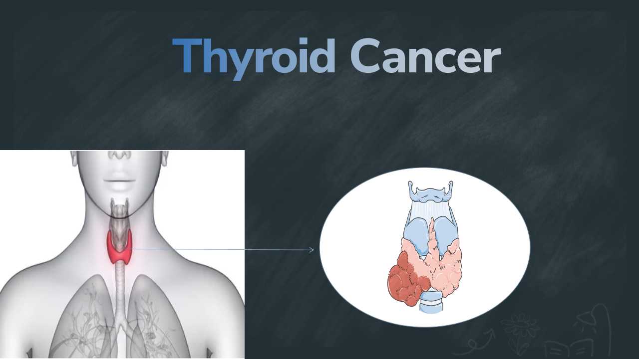
Cryptococcal meningitis
What is Cryptococcal meningitis?
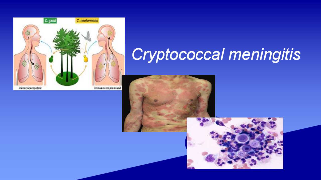
The membranes covering the brain and spinal cord are called meninges, and meningitis is an infection and inflammation of these membranes. Numerous microorganisms, such as viruses, fungi, and bacteria, can cause meningitis. Cryptococcal meningitis (CM) is caused by two different forms of fungi. Cryptococcus gattii (C. gattii) and Cryptococcus neoformans (C. neoformans) are the names given to them. Healthy individuals rarely have this infection. AIDS patients and other individuals with weakened immune systems are more likely to get CM. C.neoformans and C.gattii is a microscopic fungus that are present in the environment it is transmitted by inhalation of spores and causes crytococcosis also called cryptococcal meningitis.Most of CM are caused by a fungus known as C.neoformans. It occurs in the soil that has been exposed to bird droppings.
CM is also caused by C. gattii. It's not present in bird poop. It is found on trees, mainly eucalyptus trees. It grows in the trash that surrounds the eucalyptus tree's base. In the blood, the meningococcal meningitis germs target the cells around the capillaries, make blood leaks and capillary damage result from injury to these cells. The result of this is pink, red, and purple spots on the skin are shown. The patches could seem like small pinpricks. The rash may become more noticeable as the infection becomes worse and spreads. The dots will enlarge and get darker. It could be more difficult for people with darker skin to detect a meningitis rash. The insides of the mouth and the palms of the hands are lighter skin types that are more susceptible to rash symptoms.
Symptoms
- Fever
- Hallucinations
- Headache
- Mental status change (confusion)
- Nausea and vomiting
- Sensitivity to light
- Stiff neck
- Skin rash
- Itching
Causes/Risk factors
- Fungi(C.neoformans C.gattii)
- Enviromental exposure
- Mining
- Construction
Diagnosis
Microbiology Department
The cryptococcocal meningities is diagnosed by the microbial culture method. The sputum sample, or blood, CSF, was used as a specimen. Firstly, it grows in the cultural medium(has nutrients that help to grow) after the incubation period. Take a loopfull culture and make a slide for microscopy examination. Stain the slide with positive mucicarmine or Masson-Fontana stain.After the slide is ready, then perform microscopy examination. Microscopy examination can aid in identification of causative agent.

Cryptococcosis under the microscope
Reference:
1. https://en.wikipedia.org/wiki/Cryptococcosis
2. https://www.healthline.com/health/meningitis-cryptococcal


0 comments