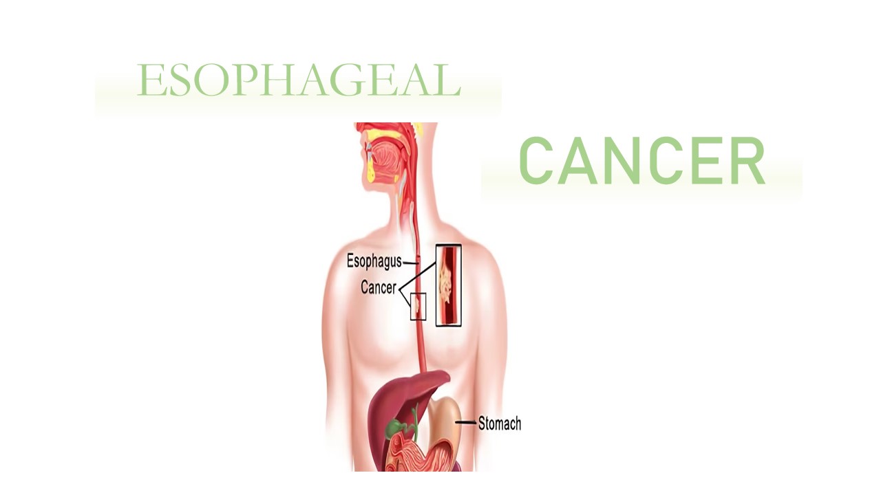
Esophageal cancer
The disease known as esophageal cancer arises when cancerous cells start to grow in the esophageal tissues.The muscular, hollow tube that transports food and liquids from the throat to the stomach is called the esophagus. Multiple layers of tissue, including muscle, connective tissue, and mucous membrane, make up the esophageal wall. As it develops, esophageal cancer moves from its initial location on the inner lining of the esophagus to its outer layers.
The types of cells that develop into malignant (cancerous) forms give rise to the names of the two most prevalent types of esophageal cancer: Cancer that develops in the thin, flat cells lining the inside of the esophagus is known as squamous cell carcinoma. Despite having the potential to develop anywhere throughout the esophagus, this cancer is most frequently discovered in the upper and middle segments. Another name for this is epidermoid cancer. Cancer that starts in glandular cells is called adenocarcinoma. Fluids like mucus are produced and released by glandular cells found in the lining of the esophagus. Usually, adenocarcinoma develops in the lower esophageal region, close to the stomach. In the world, esophageal cancer is ranked eighth. The disease is diagnosed by a biopsy with the help of an endoscope.
Symptoms
- Painful or difficult swallowing
- Weight loss
- Pain behind the breastbone
- Hoarseness and cough
- Indigestion and heartburn
- Heartburn
- Vomiting or coughing up blood
Cause/Risk factors
- Tobacco use
- Alcohaluse
- Obesity
- Barrett’s esophagus
- Chronic reflux
- Human papillomavirus(HPV)
- History of cancer
- Occupational exposure to certain chemicals
Diagnosis
Histology Department
A thin, flexible tube with a camera (endoscope) is inserted through the mouth and into the esophagus. During this procedure, small tissue samples ( known as biopsies) can be collected using specialized instruments. The collected tissue samples are fixed in formalin, preserving their structure. After fixation, the tissues are embedded in paraffin wax to create thin sections for microscopic examination. Sections of the tissue are cut and mounted on slides. Staining techniques, such as Hematoxylin and Eosin (H&E), are used to highlight cellular structures and examined under a microscope.

Squamous cell carcinoma under microscope
References:
1.https://en.wikipedia.org/wiki/Esophageal_cancer.
2.https://my.clevelandclinic.org/health/diseases/6137-esophageal-cancer


0 comments