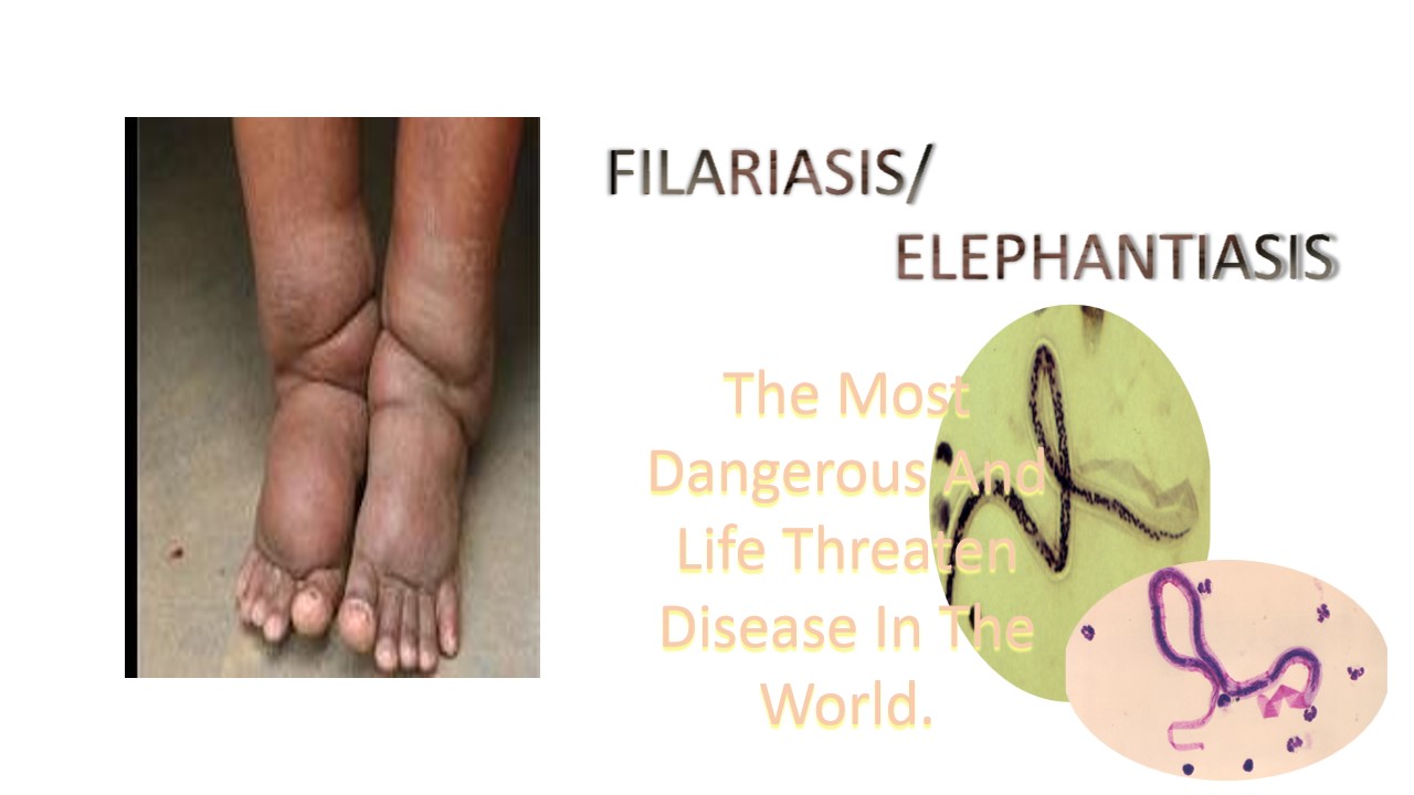
Filariasis
Filariasis is a disease caused by parasitic worms, it is transmitted to people by black files or mosquitos The parasite is a thread-like nematode, that is belong to the round worm superfamily filarioidea..The life span of filaria is 5 to 7 years. . Filariasis is also called elephantiasis disease. Filarial worms mostly affect the legs in which legs look like elephant's legs.There are three main types of parasitic worm that are cause the falariasis such as Wuchereria Bancrofti, Brugia Malayi and Brugia Timori.These worms affect the lymphatic system of the body.The lymphatic system is a network of vessels through which lymph drains from the tissue into the blood.
Mosquito bites already infected person and go and bite the other healthy person. Then the larva goes into the bloodstream of the healthy host and multiplies. In the lymphatic system, larva are nourish and become to adult filarial worm. After the became matured, the adult filarial worm release larva called microfilaria. The newly form microfilaria again enter into blood through the mosquitoes along with the host cells and repeat the cycle again and again. There are hundreds number of filaria worm discovered but now eight type of filaria nematodes are knows.The parasitic disease is a categories based on which part of the body is infected.
Lyphatic filariasis: the filaria worm(Wuchereria bancrofti, Brugia malayi and Brugia timori) damage the lymphatic system.
Subcutaneous filariasis: In which the body mostly swollen, the bottom layer of the skin and white part of the eye are infected by the Loa loa, Onchocerca volvulus, Mansonella streptocerca, and Dracunculus medinensis
serous cavity: Mansonella perstans and Mansonella ozzardi are attack abdomen serous cavity and infectedit .
Symptoms:
- Swelling of the body parts ( legs, scrotum, breast, arm)
- Fever and chills
- Limb or genital swelling
- Skin exfoliation
- chronic swelling
Causes/Risk factor
The following are the parasitic worm that causes the filariasis disease transmitted through mosquitoes.
- Wuchereria Bancrofti
- Brugia Timori
- Brugia Malayi
Diagnosis:
Hematology department
Hematological studies provide information regarding blood related diseases. The hematologist make the peripheral blood smear on glass slide and find the abnormalities or blood parasite (filaria). In the lab, the hematologist made a smear slide, fixed with methanol and stained it. In the lab, Wright’s stain, Giemsa stain and sometimes combination of both are used to diagnose the disease. Note that, collection of specimen (blood) should be drawn at night(10PM-2AM). for the identification of the filaria to diagnosed the microfilaria in the blood smear by the microscopic examination.

Microfilaria are observed in the blood smear. Purple dote indicates the nuclei inside the worm.
References:
- https://rarediseases.org/rare-diseases/filariasis/
- https://my.clevelandclinic.org/health/diseases/elephantiasis


0 comments