Kidney cancer
A class of tumors that originate in the kidney is called renal cancer or kidney cancer. The abnormal proliferation of cells in kidney tissue is known as kidney cancer. A tumor is a mass that develops from these cells over time. When a change in the cells occurs and they start to divide uncontrollably, cancer starts. A malignant tumor has the potential to spread to other tissues and important organs. This process is known as metastasis. The age range of 65 to 74 is when kidney cancer most frequently affects individuals.In youngsters, kidney cancer is far less common.
Kidney cancer comes in a variety of forms, including, in adults, renal cell carcinoma (RCC) is the most prevalent kind of kidney cancer. Clear cell renal cell carcinoma is the most prevalent kind of RCC (ccRCC). It can affect both kidneys, renal cell carcinoma often begins as a solitary tumor in one of them. The kidney's tubules, which are tiny tubes that replenish blood with nutrients and hydration, are where the cancer starts. Transitional cell carcinomas are approximately 6% to 7% of all kidney cancer. Usually, this cancer starts where the ureter joins the major portion of the kidney(this region is called renal pelvis). Additionally, transitional cell carcinoma might develop in the bladder or ureters. The rarest type of kidney cancer is renal sarcoma. If left untreated, it can spread to surrounding organs and bones from its starting point in the connective tissues of the kidneys. The most prevalent kind of kidney cancer in children is Wilms tumor. There are many methods available to diagnose kidney cancer. Biopsy is one of the methods to address kidney cancer. Which helps doctors with medication and treatment.
Symptoms
- Blood in pee (hematuria)
- A lump or mass in your kidney area
- Flank pain
- Tiredness
- A general sense of not feeling well
- Loss of appetite
- Weight loss
- Low-grade fever
- Bone pain
- High blood pressure
Causes/Risk factors
- Smoking
- Obesity
- High blood pressure
- Famiy history
- Radiation therapy
- Gene changes
- Tuberous sclerosis complex
- Von Hipple-Lindau disease(VHL)
Diagnosis:
Cytology examination
urine specimens is used to diagnose renal cancer. The urine is examined under the microscope to detect the cancerous cells. cytopathological examination indicate the abnormal cells present in the cytology specimen.

Urine cytology in detection of kidney cancer
Histology examination
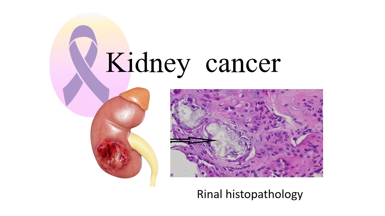
A little tissue sample from the kidney is removed during a biopsy for kidney cancer in order to be examined under a microscope. Usually, this process is carried out to identify the type and grade of the malignancy, confirm its existence, and inform treatment. The specimens is obtained from the patient and sent to the laboratory for histopathologycal examination. The pathologist fixed the sample in formalin and performed the further process also applying stain on the specimen. Staining is the crucial step, which helps to identify abnormalities and arrangement of the cells.

Renal cell carcinoma with H&E stains examined under a microscope
References:
- https://en.wikipedia.org/wiki/Kidney_cancer#Biopsy
- https://my.clevelandclinic.org/health/diseases/9409-kidney-cancer-overview


0 comments