Leptospirosis
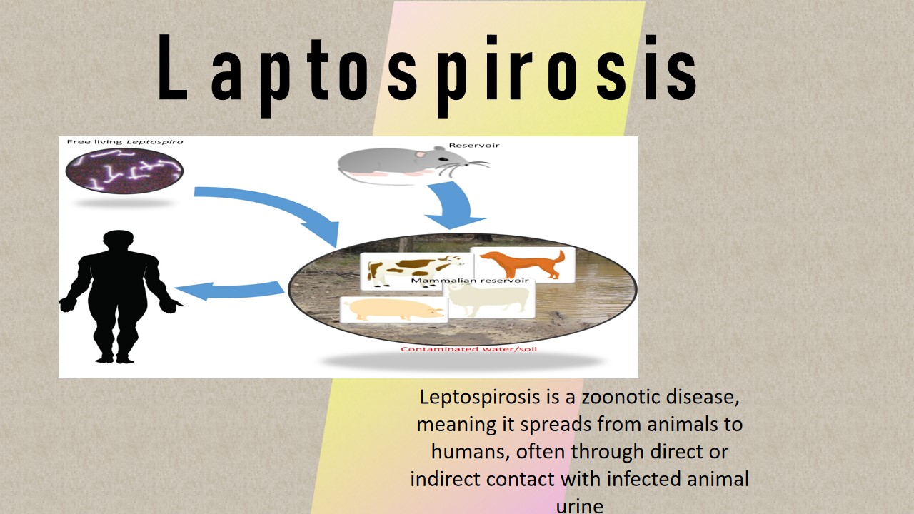
Leptospirosis also known as Weil disease that is the most prevalent zoonotic infection. An infection developed by spirochete bacterium Leptospira called leptospirosis. The size of Leptospira is found between 8 to 20µm. The most common way for it to spread is by direct contact with infected animals' urine or by coming into touch with urine-contaminated water or soil. Leptospirosis is commonly spread by farm animals like cattle, pigs, and horses, but it can also be spread by wild animals like raccoons and porcupines as well as domestic dogs.
Although leptospirosis is a global disease, it is more prevalent in tropical and subtropical regions with heavy rain weather. In Australia, northeastern New South Wales and Queensland have the highest rates of leptospirosis. Mucous membranes, such as those in the mouth and eyes, or open wounds are entry points for bacteria into the body. After that, it enters the bloodstream and travels all over the body. Anicteric syndromes and Icteric syndromes, these are two types of leptospirosis. In the anicteric syndromes, the symptoms flue like illness, it is also called first phases leptospirosis. Iciteric syndromes in which people get sick again and again. It is the second phase of syndrome or Weil disease.
Symptoms
- Fever
- Chills
- Headheach
- Vomitting
- Diarrhea
- Abdominal pain
- Cough
- Sore throat
- Jaundice
- Breathing problems
Causes/Risk factors
- infected animals
- the urine of infected animals
- contaminated soil or water
- live in tropical or temperate climates
- work with animals, like dairy farmers or veterinarians
Diagnosis
 Hematology Department: in the laboratory, the pathologist prepared a blood smear and stain with the Giemsa stain and examine under the microscope.
Hematology examination
 Cytology Department: The urine sample examine under the microscope to observed the spirochete bacterium Leptospira.
Spirochetes in urine sample
 Microbiology Department: The urine sample is obtained from the patient. For the urine sample, a centrifuged sediment is used to prepare a smear on a glass slide. The slide is then stained with Giemsa stain, which contains azure eosin dyes. After the preparation is completed, then observed under a microscope. The microscopy examination found the information about leptospira bacteria that appear as thin, tightly coiled spirochetes with hooked ends.
Thin, tightly coiled spirochetes observed under a microscope
Reference:
1.https://www.betterhealth.vic.gov.au/health/conditionsandtreatments/leptospirosis#how-does-leptospirosis-spread
2.https://www.sprintdiagnostics.in/hyderabad/test/leptospira-detection-by-dark-field-microscopy


0 comments